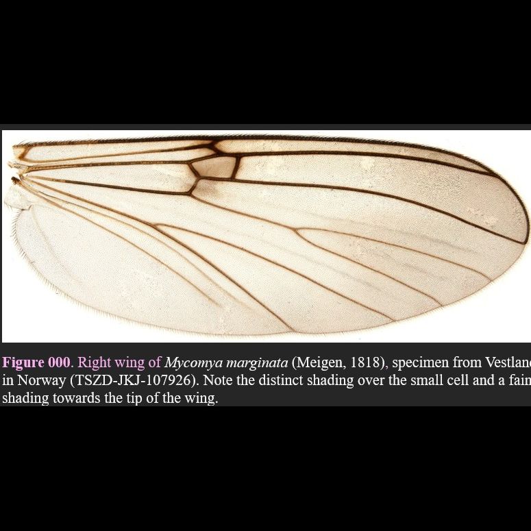

Right wing of Mycomya marginata (Meigen, 1818) (TSZD-JKJ-107926).

Note the distinct shading over the small cell and a faint shading towards the tip of the wing.
Collected from: Vestland in Norway (TSZD-JKJ-107926).
Deposited at: TMU
Identified by: JK
Adapted from:
Large species, wing length 4.61-5.89; 5.21 mm (n = 5), usually with a rather distinct coloration pattern where abdominal segment 6-7 are blackish contrasting to yellow tergites 1-5 with brown dorsal stripe. Distinguished from all other species of the M. marginata group, except M. marginella sp. nov., by having stout and large processus of tergite 9 which is broad basally and tapers into a truncated tip without any notch, with distinct brushes of stiff setae apicolaterally (Fig. 000 A); in combination with large, auriculate tergal lateral appendages (Figs 000). Separated from M. marginella sp. nov. by the structure of aedeagal apparatus which has apially veakly split but not downbent parameres and less bulging base (Figs 000 D-E). Tergite 8 narrow medially, with large, subrectangular corners armed with row of numerous seate and tapering lateral margins (Fig. 000 F). Tegal lateral appendage auriculate with rather thin setation on both sides (Fig. 000 A, C). Tergite 10 entirely reduced. Lateral corner of sternal synsclerite prolonged into medium long, very slender lateral lobe, apically setose (Figs 000 B-D). Sternal submedian appendages short, apically tapering and setose (Fig. 000 B-C). Gonostylus bilobed, lateral branch smoothly conical with slightly bent apical spine, mesial branch broad and flattened, slightly shorter, with short subapical spine and three tiny setae subapically (Fig. 000 B-C). Aedeagal apparatus shaped as typical for the species group, with balls-shaped base prolonged into sclerotized and raptor-beak-shaped parameres widening into hyaline lateral flaps preapically, without dentations in M. marginata (Figs 000 A, D-E). Double vas deferens merging to single postvesicular vas deferens shortly before entering the ejaculatoty duct ventrally on aedeagal apparatus (Fig. 000 E).
Source:
Meigen, 1818
testing
Sciophila marginata Meigen, 1818: 249 Mycomya (Mycomya) marginata – Väisänen 1984:232, figs. 1, 57, 762-768, key p.33, 41, 42 Designated as the type species for the genus Mycomya by monotypy (Väisänen, 1984)
Source:
Väisänen 1984
Mycomya marginata
First described by Meigen (1818) based on now lost type materials from Austria. We base its interpretation and taxonomic history, which is not repeated here, on Väisänen (1984). Previous records likely partly mixed with M. marginella sp. nov.
Source:
(meigen, 1818)
Palaearctic (Europe): widely distributed
Source:
multiple sources
Nordic-Baltic occurrence and national redlisting Nordic-Baltic country records. NO SE SF - KR ES LI LA DK - -. Common and widespread throughout the mainland region, most records are from southern parts of the country. Southern in NW Russia. Strangely enough not picked up through metabarcoding by the National insect monitoring in Norway. World Distribution Palaearctic, widely distributed in Europe, also recorded from Transcausasia (Georgia), Iran, West Siberia and Russian Far East (Väisänen 1984). GBIF records.
Source:
multiple sources
Habitat and Biology
Has been reared from various fungi, mostly bracket polypores, corticioid and jelly fungi (cf. Hutson et al. 1980 and Jakovlev 1994), also from Auricularia mesenterica (Edwards 1925), Merulius, Pleurotus, Sparassis crispa (Hutson et al. 1980, Jakovlev 2011), Naucoria (Chandler 1993a), Simocybe (Alexander 2002), and from fungoid wood (Zaitzev 1994). In Czech Republic the species has been reared from tussocks of grasses Calamagrostis epigejos (L.) Roth. (Poaceae) including a root ball with soil, about 25 x 25 cm; the authors supposed that occurrence of larvae known as web-spinners on the surface on various wood decaying fungi in this case could be accidental (Ševčík & Roháček 2008). Finnish and Russian specimens are chiefly from natural and seminatural boreal forests, several records also from burnt clear-cuts. Adults collected with sweet baits, Malaise and light traps. In Germany obtained with emergence traps over beach (Fagus sylvatica) stumps (Irmler et al. 1996); in Southern Finland by emergence traps on slash residues of birch and pine on clear-cuts.
Source: